Onion Cell Diagram
Onion microscope drawing labelled px biological Onion cell hi-res stock photography and images Biopedia: practicals
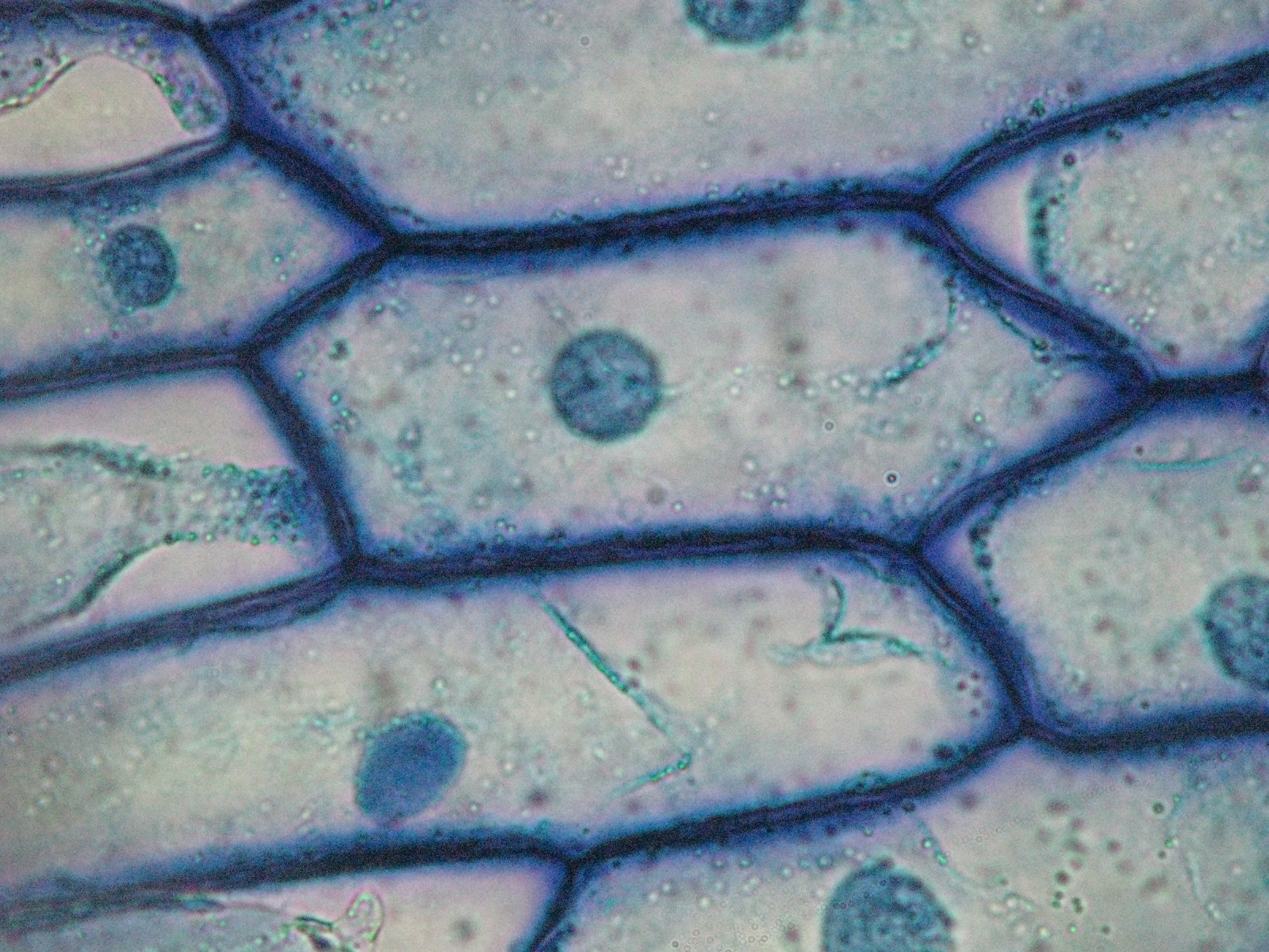
Beautiful World: Onion cells
Can someone please post me a diagram of Onion cells 2 Onion cell microscope micrograph cells microscopic section allium cepa scale epidermis alamy root bulb tip organelles
Onion cells cell diagram biology help microscopic structure occupies uses introduction
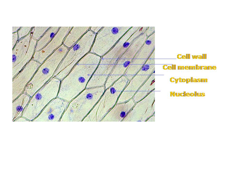
Onion epidermal cell labeled diagramOnion cell diagram drawing Label cells procedureOnion cell diagram labeled epidermal wiring.
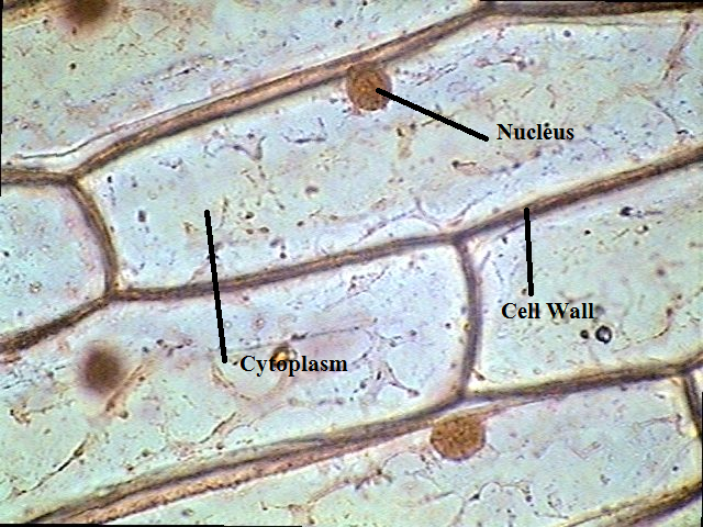
Draw a well labelled diagram for the cell observed in onion peel andOnion cell epidermal labeled diagram cells microscope under drawing skin epidermis lab bulb nucleus membrane mag preparation cytoplasm vacuole leaves Cells microscope labelled stainStaining of onion cell nuclei – microbehunter microscopy.
Onion cell epidermal peel size
Onion cell under microscope labeled drawingOnion labelled cheek observed Onion bulb structure allium cepa foods red bulbs schematic figureThe science scoop: onion cell lab.
Onion peelOnion cell diagram epidermal labeled cells leaves modified bulb formed microscope under Fun science blog: onion cellBeautiful world: onion cells.

Magnified times 100x microscopy
Dissection labeled organellesOnion epidermal cell diagram Onion epidermal cell labeled diagramOnion cell 400x lab microscope under labeled cells structure scoop science looked.
Onion nucleusOnion cells under microscope Onion epidermal cell labeled diagramGordon cooke » student images and videos from the microscope practical.

Onion cell cells lab nucleus staining nuclei microscope under stained skin dna simple look experience class there students tissue microscopy
Biology help online: learning on onion cellsOnion peel cell diagram with label Cell onion plant presentation cheek slideserve ppt powerpoint slide1Onion cells beautiful.
Epidermal labeled epidermis biology chromosome observationOnion peel cell diagram with label Onion cells.
![Can someone please post me a diagram of - 1] Onion cells 2] Animal](https://3.bp.blogspot.com/_f_OmAdlU5os/Sq0MoOl7_cI/AAAAAAAAAqs/AooDOMrMUUY/s400/scan0007+-++2.jpg)
Biopedia: Practicals

Fun Science Blog: Onion Cell

Onion Cells under Microscope

Onion Epidermal Cell Labeled Diagram - Wiring Diagram Pictures

PPT - Onion Cell PowerPoint Presentation, free download - ID:1962002

Onion Epidermal Cell Labeled Diagram - Wiring Diagram Pictures

Staining of Onion Cell Nuclei – Microbehunter Microscopy
The Science Scoop: Onion Cell Lab